
Abstract Article historyThe odontogenic keratocysts are developmental cysts of the jaws that require proper diagnosis due to their potential for. These diagnostic tools allow the dental professional to identify the type of cyst and plan appropriate treatment.

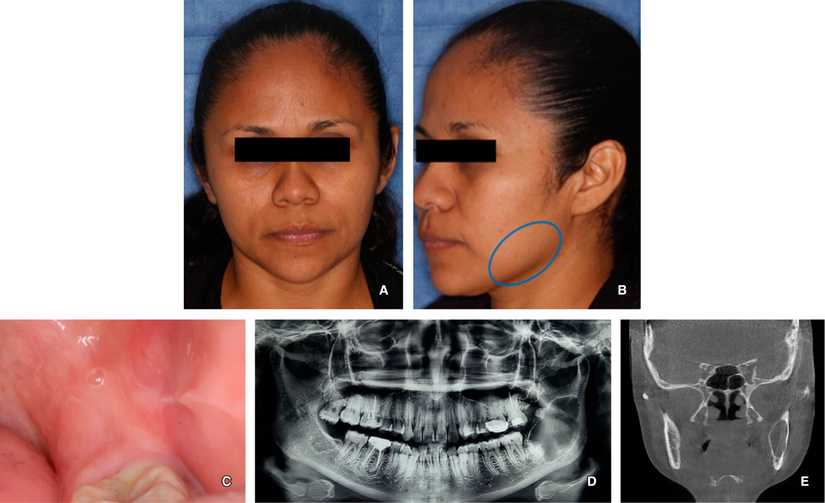
Case Presentation Of A Patient Affected By Okc That Didn T Experience Download Scientific Diagram
Differential diagnosis Imaging differential considerations include.

. Journey of okc from cyst to tumor to cyst again. Implant surgery for the placement of dental implants is performed after full bony consolidation of the bone grafts to complete full oral rehabilitation for the patient. Biopsy results came back positive with the diagnosis of odontogenic keratocyst in the left mandibular region.
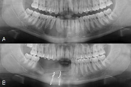
Multiple surgical approaches were introduced including decompression marsupilization. Incision on the right mandibular ramus up to the distal aspect of tooth from the buccal aspect. The cyst was enucleated and the infected bone was debrided.
The most appropriate surgical approaches for the successful. Nancy Herbst walks through how to properly remove an Odontogenic KeratocystLike and subscribe for moreUnion City Oral Surgery Group is located in Union. Case Report Figure 4.
Dental professionals will typically recommend a test like an MRI CT or X-ray. Odontogenic keratocyst are surgically removed by marsupialisation enucleation and chemical curettage with carnoys solution. In rare instances particularly large cysts may require resection and bone grafting.
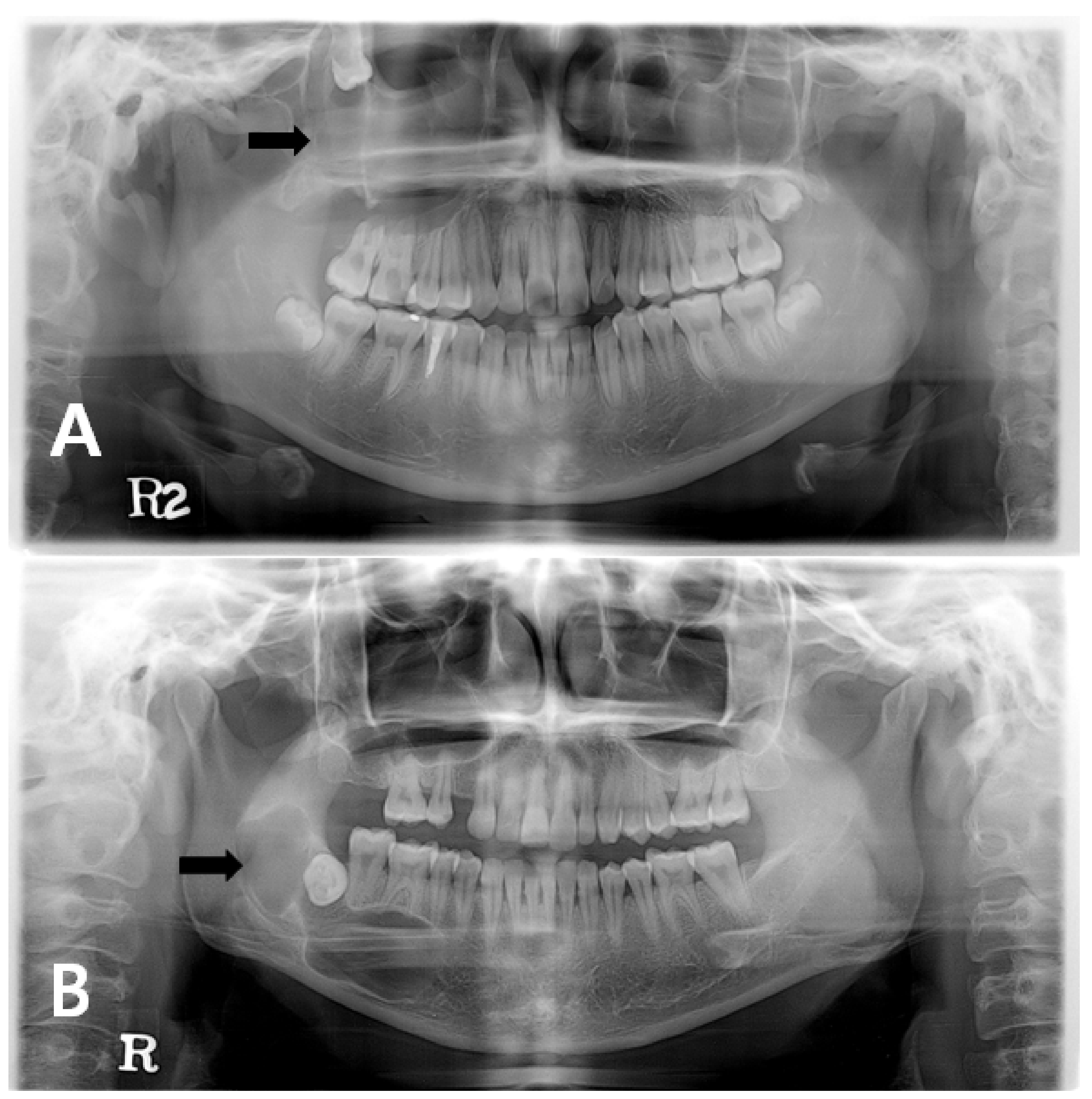
There was a recurrence of the cyst in a few months and. Odontogenic keratocyst okc or keratocystic odontogenic tumor kcot. Large odontogenic keratocysts sometimes are treated initially by cystotomy and insertion of a drainage tube which can promote shrinkage of the lesion and fibrous thickening of the cyst wall before subsequent total removal.
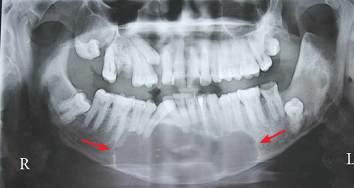
CT scans in axial and coronal planes. Long-term follow-up with monitoring by X-ray is important as if these cysts are left untreated they can become quite large and locally destructive. We recommend the following protocol in the management of large mandibular OKC.
Treatment planning was explained to him and he consented to surgery. Odontogenic keratocysts make up around 19 of jaw cysts. Treatment of the odontogenic keratocyst involves meticulous resection to completely remove the lesion followed by reconstruction of the jaw with bone grafting.
In the WHOIARC classification of head and neck pathology this clinical entity had been known for years as the odontogenic. Dentigerous cyst both dentigerous cysts and odontogenic keratocysts can be positioned pericoronally dentigerous cysts tend to attach at the cemento-enamel junction of teeth radicular cyst. Comprehensive review with recent updates on who classification 2017.
Enucleation of the cyst and removal of the associated teeth. It was first described by H P Philipsen in 1956 as an odontogenic keratocyst. Download Citation Removal of Odontogenic Keratocyst in Maxilla Through the Le Fort I Osteotomy The odontogenic keratocyst is a lesion with specific clinical and histopathological aspects.
An odontogenic keratocyst is a rare and benign but locally aggressive developmental cyst. It is advantageous for complete removal without the risk of damage to the ossification centers of. The principle of treatment of odontogenic.
Surgical Removal of Odontogenic Keratocyst. Odontogenic keratocysts OKCs are benign intraosseous odontogenic lesions that have a locally aggressive behavior and exhibit a high recurrence rate after the treatment. Odontogenic keratocysts can initially be treated with incisional biopsy and decompression by installing a polyethylene drain to allow subsequent reduction of the cystic cavity size resulting in thickening of the capsule which allows a later easy removal withapparently lower relapse rate waldron.
They may also suggest getting a biopsy where you will get part of the cyst removed and sent to a laboratory for further examination. Biopsy of the lesion. In rare instances particularly large cysts may require resection and bone grafting.
Cyst can be removed by open as well as endoscopic approach. Imaging studies and a biopsy were obtained at the hospital. Large odontogenic keratocysts sometimes are treated initially by cystotomy and insertion of a drainage tube which can promote shrinkage of the lesion and fibrous thickening of the cyst wall before subsequent total removal.
The excision of the overlying mucosa. Odontogenic keratocyst is one of the most aggressive odontogenic cysts with a high recurrence rate this was explained histopathologically as it typically shows a thin friable wall which is often difficult to enucleate from the bone in one piece and have small satellite cysts within the fibrous wall. Immediate mandibular reconstruction with a corticocancellous iliac crest bone graft.
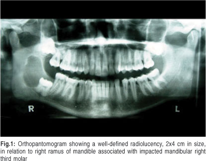
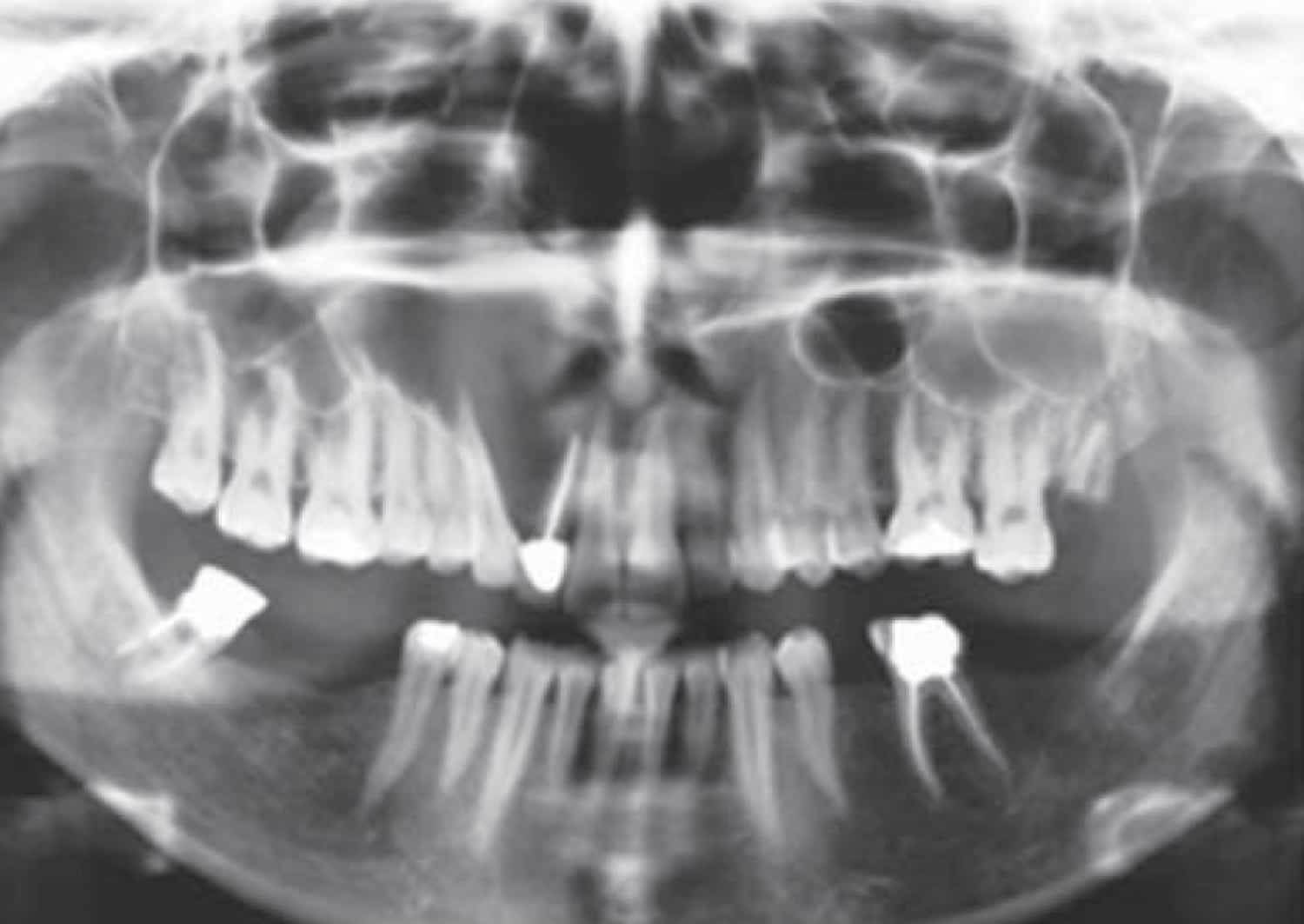
It most often affects the posterior mandible and most commonly presents in the third decade of life. Osteotomy in the trigonoretromolar region until the exposure of the lesion. Enucleation of the lesion.
Removal of the cyst with removal of surrounding bone and or cryosurgery intense cold is applied to the cyst and bone are the most common forms of treatment. Regardless of the size of the lesion or treatment modality continued clinical. First described by Philipsen in 1956 the odontogenic keratocyst is characterized by a large squamous keratinization of its border an aggressive growth and a.
Surgical enucleation of Odontogenic Keratocyst. We opted for an endoscopic approach because it offers minimal reductive change.
Management Of Recurrence Of Ameloblastoma And Odontogenic Keratocyst A Cross Sectional Study

Pdf Removal Of Odontogenic Keratocyst In Maxilla Through The Le Fort I Osteotomy Semantic Scholar

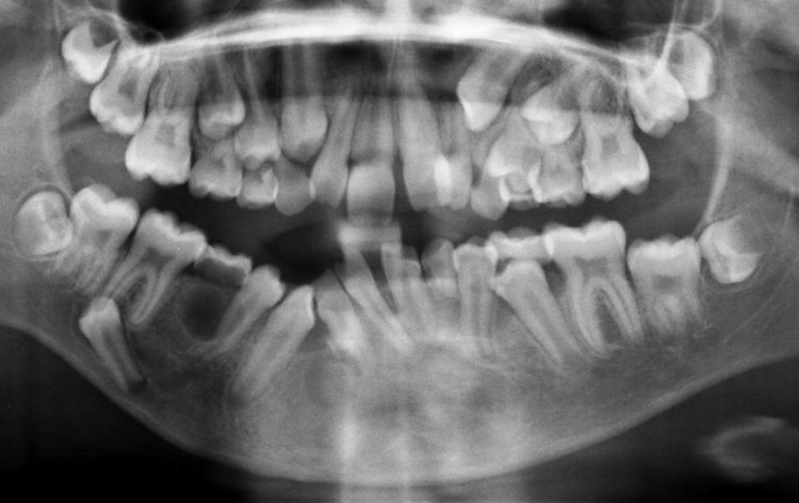
Management Of Benign Odontogenic Lesions In The Paediatric Patient
Orthokeratinized Odontogenic Cyst Critical Appraisal Of A Distinct Entity

Endoscopically Assisted Enucleation And Curettage Of Large Mandibular Odontogenic Keratocyst Oral Surgery Oral Medicine Oral Pathology Oral Radiology And Endodontics
Jcm Free Full Text Changes In Cellular Regulatory Factors Before And After Decompression Of Odontogenic Keratocysts Html
Odontogenic Keratocyst Okc Exodontia
Odontogenic Keratocyst Definition Causes Symptoms Diagnosis Treatment Prognosis
Odontogenic Keratocyst Okc Exodontia

Keratocystic Odontogenic Tumor Treatment Modalities Study Of 3 Cases

Review Of 5 Fluorouracil Is Associated With A Decreased Recurrence Risk In Odontogenic Keratocyst Management A Retrospective Cohort Study Journal Of Oral And Maxillofacial Surgery
Odontogenic Keratocyst Definition Causes Symptoms Diagnosis Treatment Prognosis

Conservative Management Of Odontogenic Keratocyst With Long Term 5 Year Follow Up Case Report And Literature Review Sciencedirect

Recurrence Of Odontogenic Keratocyst Okc In Relation To Cortical Download Scientific Diagram
Treatment With Decompression Of An Odontogenic Keratocyst

Treatment Of The Keratocystic Odontogenic Tumor Kcot In Patients With Gorlin Goltz Syndrome A Review Of The Literature With A Case Report Italian Journal Of Dental Medicine

Conservative Technique For Enucleation Of A Large Dentigerous Cyst Through Bony Fenestrations British Journal Of Oral And Maxillofacial Surgery
Odontogenic Keratocyst Okc Exodontia

Jaw Reconstruction Surgery With Bone Graft Odontogenic Keratocyst